What is the finding
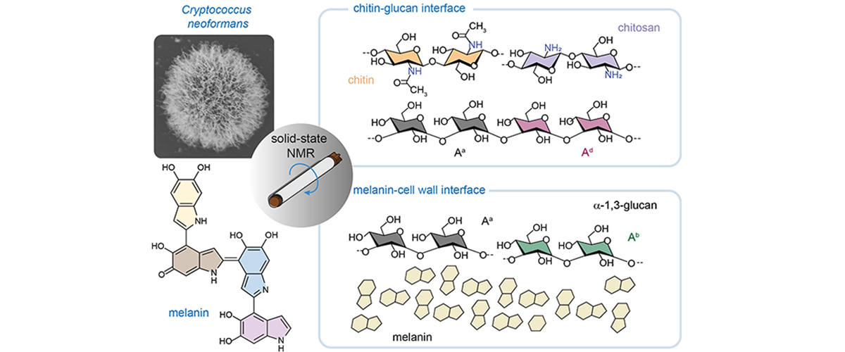
Using a powerful magnetic resonance technique called Dynamic Nuclear Polarization (DNP), scientists discovered that an invasive fungus called Cryptococcus contains five different forms of a sugar known as α‑glucan. These sugars act like internal scaffolding inside the fungal cell wall. They physically link together rigid fibers and two protective outer layers, helping hold the cell together. This sturdy framework makes the fungus stronger, better able to survive in the human body, and more resistant to antifungal drugs.
Why is this important?
Treating cryptococcosis often requires long courses of harsh antifungal drugs that are expensive and can cause serious side effects. Many newer antifungal drugs attack fungal cell walls, but they work poorly against Cryptococcus. These findings show that a key sugar in the Cryptococcus cell wall plays multiple structural roles, helping explain the fungus’s strength and why existing drugs are less effective, but also highlighting new opportunities to design better, more targeted antifungal therapies.
Who did the research?
Ankur Ankur1, Jayasubba Reddy Yarava1, Isha Gautam1, Faith J. Scott2, Frederic Mentink-Vigier2,3, Christine Chrissian4, Li Xie1, Dibakar Roy1, Ruth E. Stark4, Tamara L. Doering5, Ping Wang6, Tuo Wang1
1Michigan State University; 2National MagLab; 3FSU; 4City College of New York; 5Washington University; 6Louisiana State University
Why did they need the MagLab?
The MagLab’s powerful magnets and specialized Dynamic Nuclear Polarization (DNP) facility made it possible to detect a rare, previously unknown form of α‑glucan. These tools also revealed how this sugar interacts closely with other cell‑wall components inside living cells. This level of detail—showing how molecules are packed together—cannot be achieved with standard laboratory methods and was essential for understanding how the fungal cell wall is built.
Details for scientists
- View or download the expert-level Science Highlight, Insight into Cryptococcus Cell Wall, Capsule, and Melanin Using Solid-State NMR
- Read the full-length publication, Polymorphic α-Glucans as Structural Scaffolds in Cryptococcus Cell Walls for Chitin, Capsule, and Melanin: Insights From 13C and 1H Solid-State NMR, in Angewandte Chemie International Edition
Funding
This research was funded by the following grants: K. M. Amm (NSF DMR-2128556); Wang (NIH AI173270); T.L. Doering (NIH AI192892; AI175875); R.E. Stark (NIH AI171093); NHMFL (NIH GM148766)
For more information, contact Zhehong Gan.