What did the scientists discover?
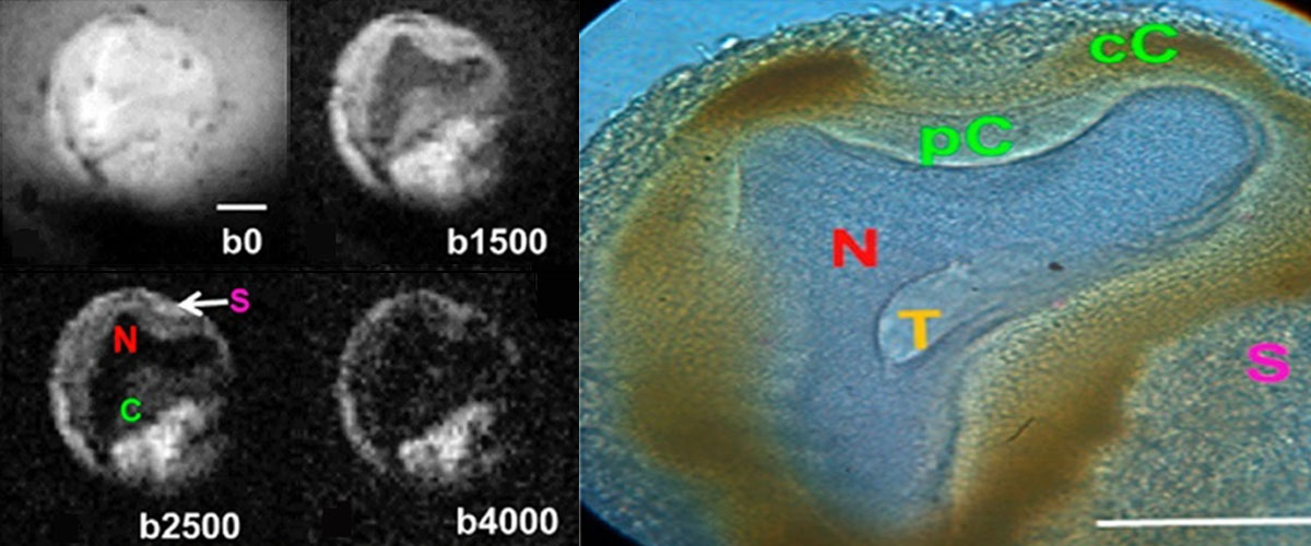
Magnetic resonance imaging (MRI) is a leading diagnostic technique for neurological studies. Glial cells in the brain are responsible for maintaining water balance to ensure accurate and consistent neuron function. In MRI scans taken within the first hours after a stroke, the images appear bright around the injury, but this has been a mystery because the direct effect on neuronal swelling appears dark. In this work, a MagLab user collaboration finds that it is the changes in glial cell volumes that cause the bright effect in the MRI signal after stroke.
Why is this important?
This work is a milestone of thirty-five years of ongoing research, starting with the first MRI image of a single cell by the lead collaborator on this team. A decades-long-held belief had been that MRI image changes seen in stroke were due to neuronal swelling. Instead, the finding is that it is glial cell swelling, leading to a better understanding of the changes in MRI signals that might one day distinguish reversible and irreversible stroke events that require different treatments and may provide better diagnostics for a myriad of other brain disorders.
Who did the research?
SJ Blackband1,2, JJ Flint1, B Hansen3 , TM Shepherd4 , CH Lee1,4, WJ Streit1, JR Forder1,2
1University of Florida; 2National MagLab; 3Aarhus University, Denmark; 4New York University School of Medicine
Why did they need the MagLab?
Physicists and neuroscientists utilize highly sensitive MR imaging gradients and microcoils at the AMRIS Facility because they enable resolution of perfused neuronal cells and capture diffusion measurements at the microscopic cellular level. Instrumentation is being developed to improve these measurements by moving to higher magnetic fields, including the MagLab's unique 32T superconducting magnet.
Details for scientists
- View or download the expert-level Science Highlight, On the Origin of MRI Signal in Stroke
- Read the full-length publication, On the Origins of Diffusion MRI Signal Changes in Stroke, in Frontiers in Neurology.
Funding
This research was funded by the following grants: Boebinger (NSF DMR-1644779); Blackband (NIH R01EB012874)
For more information, contact Joanna Long.