What did the scientists discover?
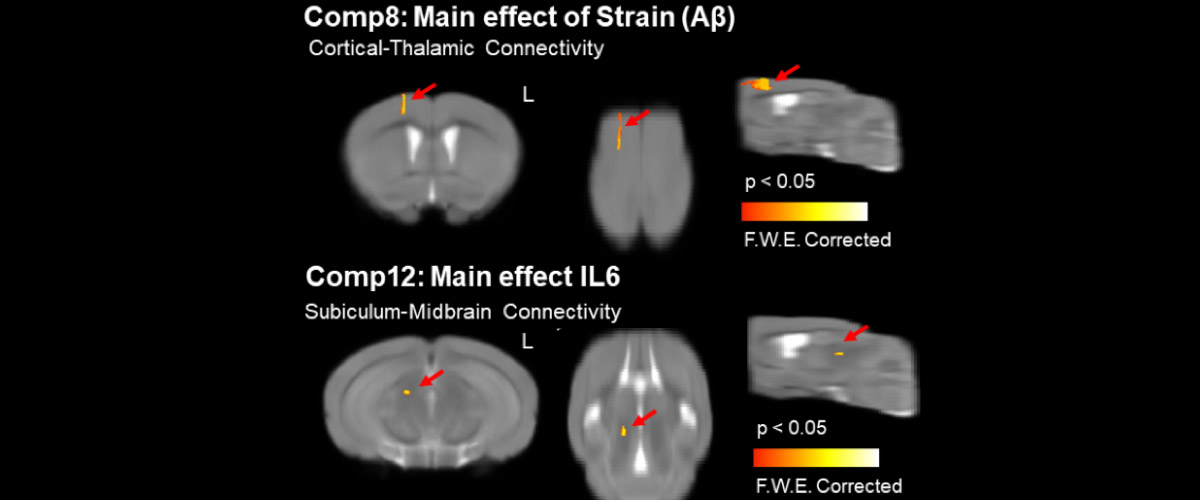
Magnetic Resonance Imaging (MRI) can be used to measure changes in brain microstructures of mice affected by Alzheimer's disease, an inflammatory brain disease with effects that disrupt the limbic functions of emotion, learning, and memory.
Why is this important?
Alzheimer's disease is a neurodegenerative condition linked to plaque deposits in the brain. The study of mouse models of Alzheimer’s disease can be used to determine the roles of extracellular β-amyloid (Aβ) plaque deposits and inflammation in affecting brain morphology and function. Quantifying changes in the brain using MRI will help to monitor and predict disease progression, as well as potentially suggest new treatment methods.
Who did the research?
L. Colon-Perez1,4, K. R. Ibanez2,5, M. Suarez4, K. Torroella4, K. Acuna4, E. Ofori1,6, Y. Levites1,2,5,7, D. E. Vaillancourt1,3,4,6, T. E. Golde1,2,5,7, P. Chakrabarty1,2,5,7, M. Febo1,3,4,7
1Florida Alzheimer's Disease Research Center, 2Center for Translational Research on Neurodegenerative Diseases, U. Florida; 3AMRIS Facility, 4Psychiatry, 5Neuroscience, 6Applied Physiology & Kinesiology, 7McKnight Brain Institute
Why did they need the MagLab?
Specialty MRI coils created for the unique 11.1T MRI magnet system at the MagLab's AMRIS Facility, in conjunction with the strong MRI technique experience of the AMRIS staff, enabled the acquisition of high-resolution images of mouse brains. The diffusion MRI and functional connectivity analyses were carried out at 11.1T in mice with early onset deposit of amyloid plaque in the brain.
Details for scientists
- View or download the expert-level Science Highlight, MRI detects brain responses to amyloid plaque deposition and inflammation
- Read the full-length publication, Neurite orientation dispersion and density imaging reveals white matter and hippocampal microstructure changes produced by Interleukin-6 in the TgCRND8 mouse model of amyloidosis, in NeuroImage.
Funding
This research was funded by the following grants: NIH P50AG047266, R01NS052318, R01NS075012, T32NS082168, P50NS091856, 1R01AG055798; G.S. Boebinger (NSF DMR-1157490, NSF DMR-1644779)
For more information, contact Joanna Long.


