What is the finding
This study explores how the African spiny mouse (Acomys cahirinus), a rodent known for its ability to heal damaged tissue, recovers from a stroke. Unlike previous findings in spinal cord repair, MRI scans showed the mice didn’t regrow brain tissue. Instead, they recovered quickly by using other parts of their brain to compensate for the damage.
Why is this important?
Ischemic stroke happens when a blocked blood vessel cuts off oxygen from the brain. It is a major cause of disability and death in adults over 40. This study used MRI to map the African spiny mouse’s brain and see how it adapted after injury from stoke, finding that it recovers unusually well from strokes. Understanding how this mouse rewires its brain could provide valuable insights into broader brain development and healing in mammals.
Who did the research?
Benjamin M. Kidd, Justin A. Varholick, Dana M. Tuyn, Pradip K. Kamat, Zachary D. Simon, Lei Liu, Mackenzie P. Mekler, Marjory Pompilus, Jodi L. Bubenik, Mackenzie L. Davenport, Helmut A. Carter, Matteo M. Grudny, W. Brad Barbazuk, Sylvain Doré, Marcelo Febo,Eduardo Candelario-Jalil, Malcolm Maden, & Maurice S. Swanson
University of Florida
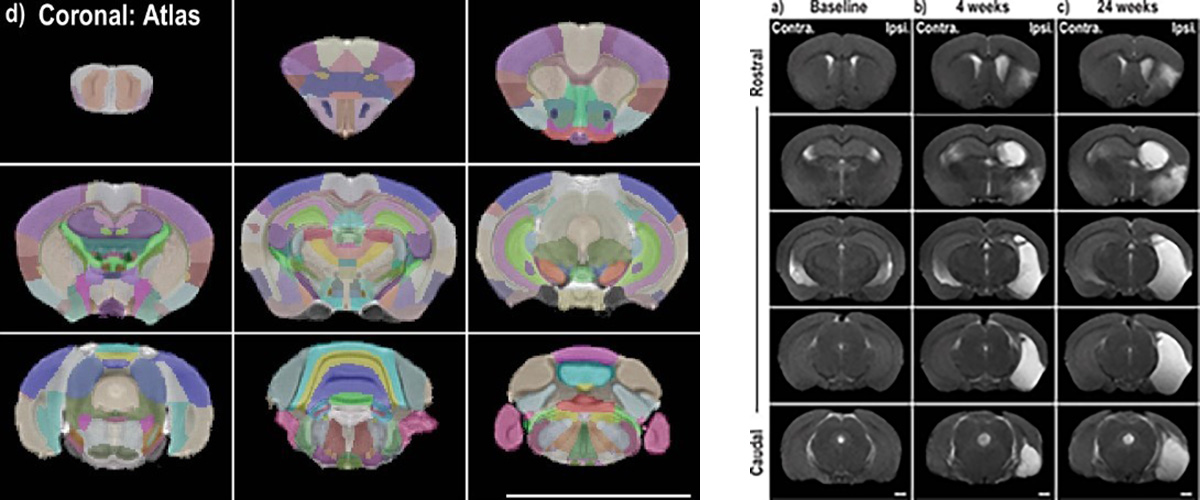
Why did they need the MagLab?
This study used advanced MRI in three ways: First, to track how the brain’s structure changed after a stroke. Second, resting-state functional MRI (rsfMRI) showed how different brain areas connect and adapt to compensate for damage. Third, detailed post-mortem MRI created a high-resolution map of the spiny mouse brain, helping researchers interpret earlier results and support future studies. These techniques required powerful high-field MRI technology, only possible with the MagLab’s specialized equipment.
Details for scientists
- View or download the expert-level Science Highlight, Stroke-induced neuroplasticity in spiny mice in the absence of tissue regeneration
- Read the full-length publication, Stroke-induced neuroplasticity in spiny mice in the absence of tissue regeneration, in Nature Partner Journals (npj) Regenerative Medicine
Funding
This research was funded by the following grants: M. S. Swanson (NIH P50 NS048843), K. M. Amm (NSF DMR-2128556; NSF DMR-1644779); A. S. Edison (NIH S10 RR025671)
For more information, contact Joanna R Long.