The MagLab is funded by the National Science Foundation and the State of Florida.
The MagLab is funded by the National Science Foundation and the State of Florida.
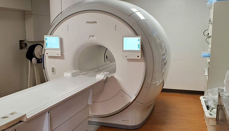
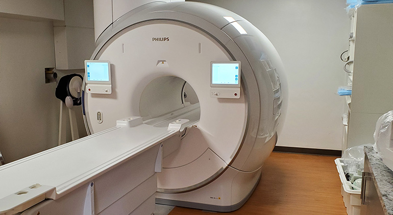
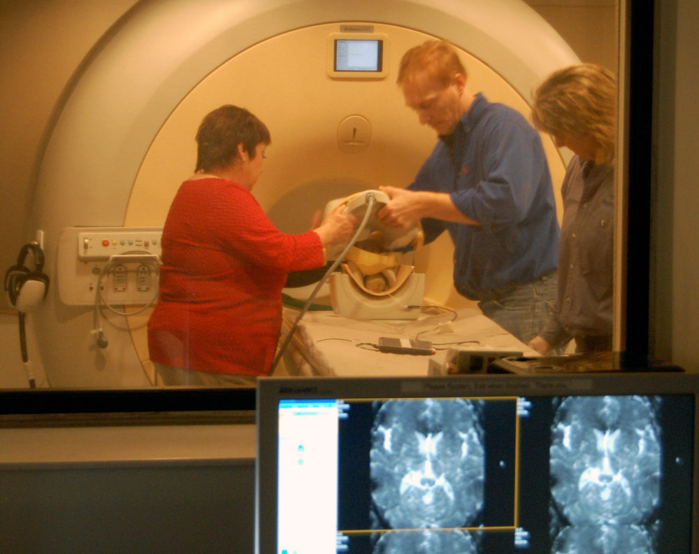
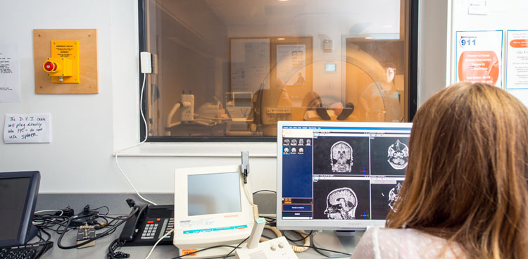
This instrument is located at the MagLab's AMRIS Facility at the University of Florida in Gainesville.
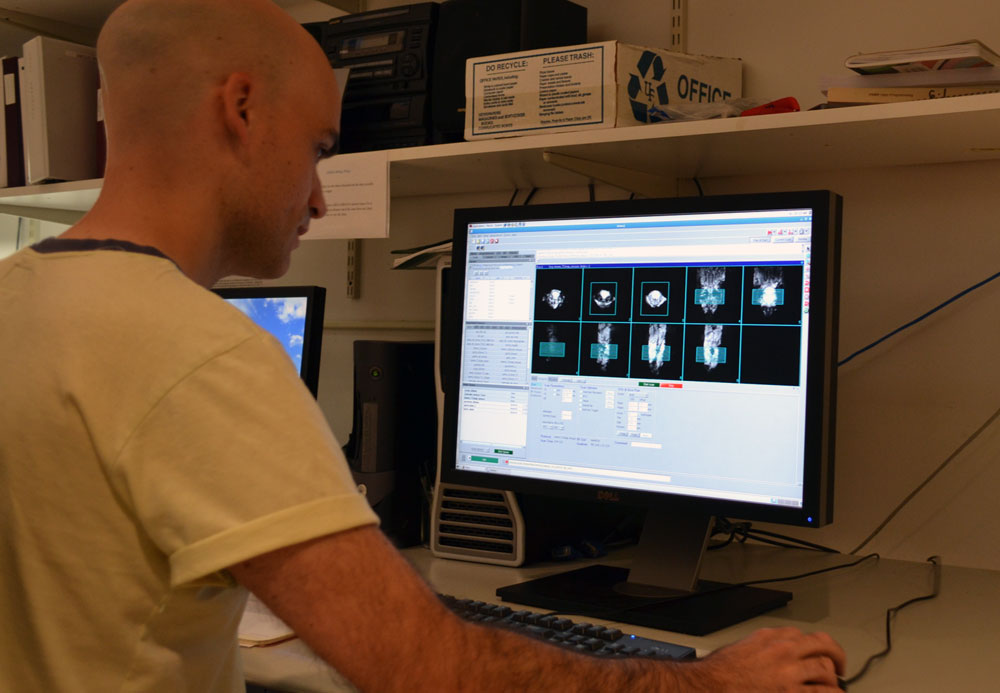
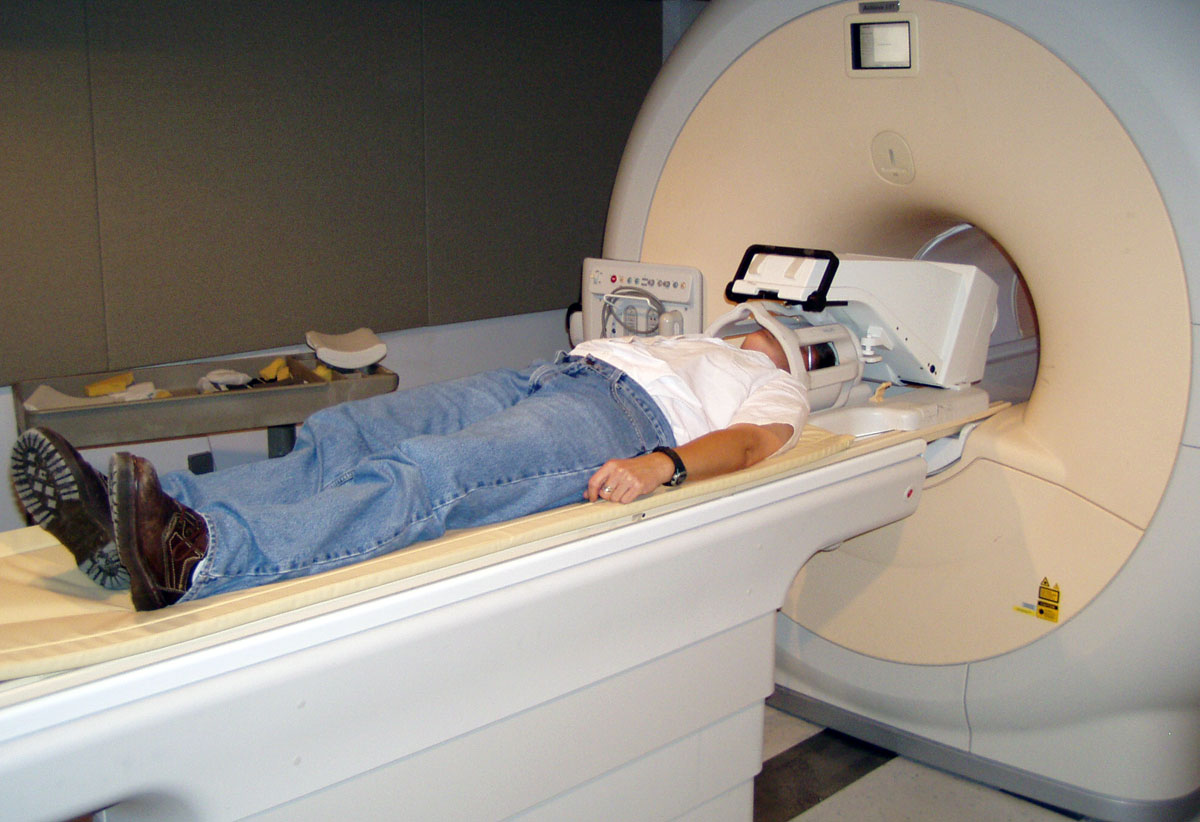
This Philips whole-body 3 T MRI scanner features a 70 cm bore and includes the latest acquisition techniques for human MRI research, including multinuclear capabilities, functional MRI (fMRI) and advanced diffusion imaging (dMRI), and magnetic resonance elastography (MRE), spectroscopy (MRI/S), whole-body scanning and much more. The extra space better accomodates in-bore equipment, like the Resoundant probe for MR elastography studies of the liver. The magnet provides the largest homogenous field-of-view, 55 cm, in a commercial 70 cm system while still providing strong gradients. The MR7700 is equipped with Philips’ unique broadband digital MR architecture, the d-STREAM, which samples the signal directly at the coil and transmits the signal through fiber optics to reduce noise and increase SNR. The system comes with the latest technologies for neuro, cardiac, MSK, liver, and whole-body imaging including, compressed SENSE technology, and numerous RF coil and pulse sequence packages for advanced MR imaging and spectroscopy research. The system is equipped with a BOLDscreen32 from Cambridge Research Systems for functional MRI task paradigms presentation as well as entertainment during MRI scan. A plug-and-play button response device ("Package 932") by Current Designs Inc. is available for collecting subject responses in fMRI studies. As of January 2023, we will expand our capabilities with a Xenon (129Xe) polarizer from Polarean for lung imaging applications. The polarizer comes with a measurement station and 129Xe coil. The polarizer is available to researchers pending project review and approval from Polarean Inc.
The following 3rd party coils are available for research use. Interested users shouldcontact Dr. Jens Rosenberg.
Explore our magnet schedule to see what exciting research is happening on our stellar fleet of instruments right now.
Credit: National MagLab
When an investigator is planning a project that will use the 3T MRI/S scanner, the investigator(s) must discuss the project with Dr. Jens Rosenberg to review their needed protocol and make sure the needed instrumentation and techniques are available, i.e. hardware exists, protocols may be put together readily, and that sufficient protocol development and/or validation time is allocated. If necessary, Dr. Rosenberg can assist in developing an imaging protocol based on specific needs of a project; it is important to realize that optimizing a specific protocol for a particular project is critical to obtaining significant data. For an ongoing project, if a new device is added and some testing is needed as planned when the project was launched, additional protocol development up to 1 hour without charge may be approved by the Director of the 3T Facility (i.e., Dr. Rosenberg).
The AMRIS Facility can allocate up to 2 hours of development time for each funded project to implement and test protocols. If the development is more involved than can be accomplished in this time, the investigator(s) must fund additional time from their project budget and/or include Dr. Rosenberg in the project as a co-investigator. For pilot studies, there are several funding mechanisms available through the MBI, CTSI, and SECIM programs at UF.
If the hardware required for a project does not exist and/or the necessary MR protocols or sequences are not currently available on the 3T, Dr. Rosenberg will discuss options for acquiring the needed hardware, sequences and/or developing the required protocol. If the needed MR techniques and protocols are not already available on our scanner and will require Dr. Rosenberg's time to develop them, the grant application budget must include Dr. Rosenberg's time as an MR physicist in addition to any funds needed for acquisition of the hardware and/or software.
Investigator(s) are also encouraged to review with Dr. Rosenberg their needs for processing and analysis of data acquired on the 3T MRI/S scanner in order to ensure sufficient resources are allocated for this purpose in grant applications.
For more information contact Jens Rosenberg, phone; 352-294-8811 or visit : https://amris.mbi.ufl.edu/mri-systems/3t-philips/
Last modified on 05 February 2026
Search our magnets by field strength, bore size, and even technique.