What did the scientists discover?
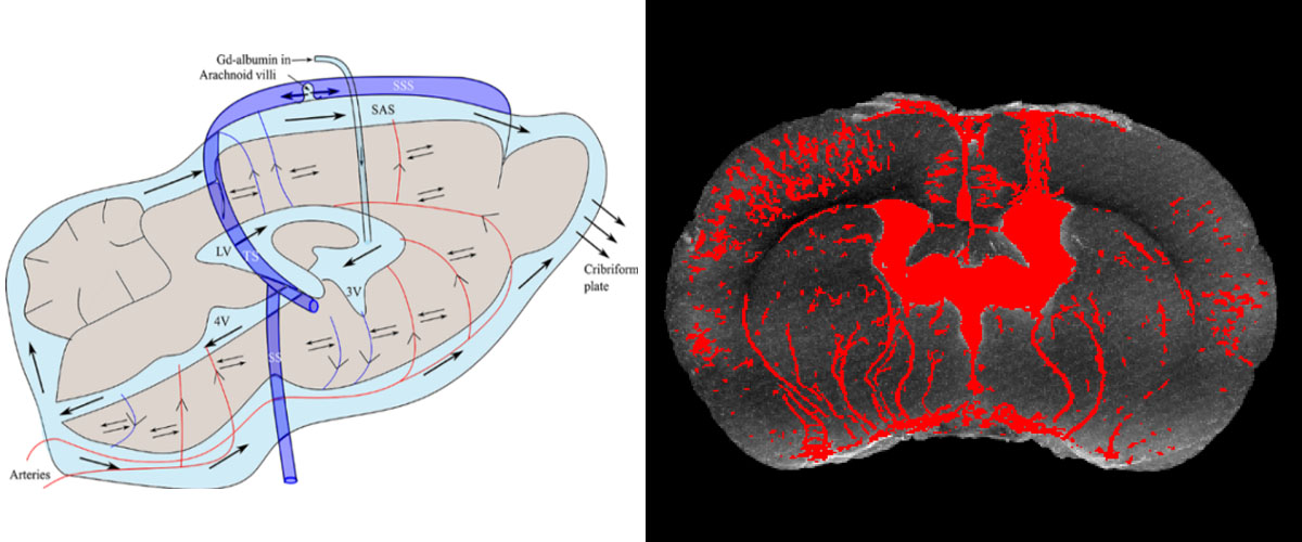
MRI can provide exquisite visualization of the brain with the resolution necessary to see the small annular spaces around cerebral blood vessels known as the peri-vascular network (PVS). Using high-field magnetic resonance imaging, researchers found a possible pathway for metabolic waste removal from the brain - a small annular space around penetrating blood vessels in the brain that opens to fluid flow and widens during sleep.
Why is this important?
This finding demonstrates a possible pathway for metabolic waste clearance from the brain with the ventricles providing a source or sink for metabolites. This may be related to the daily requirement for sleep, since recent studies indicate that the perivascular network dilates during sleep, enabling enhanced flow.
Who did the research?
Kulam Najmudeen Magdoom1, Alec Brown1, Julian Rey1, Thomas H. Mareci2, Michael A. King3 & Malisa Sarntinoranont1
1University of Florida, 2National MagLab, 3Veterans Administration;
Why did they need the MagLab?
At present, this research can only be performed at the MagLab's AMRIS Facility because it requires the use of a high magnetic field (17.6 T), wide bore (89 mm) imaging spectrometer. This instrument was specifically designed for high resolution spectroscopy and imaging (combining high magnetic field, strong gradients, unique MRI coils, and high field homogeneity) and provides the sensitivity necessary for detecting contrast-enhanced MR at the high resolution needed to visualize the three dimensional PVS network within an intact brain and find connections between the ventricles and the functional tissue of the brain.
Details for scientists
- View or download the expert-level Science Highlight, High Magnetic Field MRI Evidences Pathways for Metabolic Brain Waste Clearance
- Read the full-length publication, MRI of Whole Rat Brain Perivascular Network Reveals Role for Ventricles in Brain Waste Clearance, in Nature Scientific Reports.
Funding
This research was funded by the following grants: G.S. Boebinger (NSF DMR-1157490); K. Magdoom (Chiari & Syringomyelia Foundation
For more information, contact Joanna Long.
