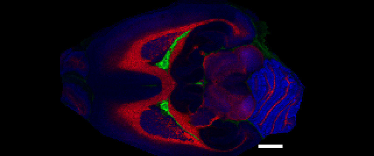
Last year, Smith teamed up with Ron Heeren from the Maastricht MultiModal Molecular Imaging Institute at Maastricht University in the Netherlands. With their team, the scientists ran a month’s worth of experiments in the 21-T, examining very thin slices of brain from healthy rats. In each 24-hour experiment, they focused on specific biomolecules. In the two data sets examined for the Analytical Chemistry article, the team looked for certain lipids, a class of biomolecules that execute critical functions in the body, including in cell membranes.
The image above, for example, shows the distribution of three specific lipids in one tissue slice. In red is phosphatidylcholine O-36:2; in green is sphingomyelin 40:1; and in blue is phosphatidylcholine 40:6. (The white bar has a length of 2 mm, approximately 5/64 of an inch.)
Mass spectrometers are fancy molecular scales that use a strong magnet to identify each molecule in a substance by its unique mass. The molecules must first be given a positive or negative charge (ionized) so that the magnet can detect them. The team used a technique called matrix-assisted laser desorption ionization (or MALDI, used for the first time on the 21-T for this project) to methodically vaporize, ionize and measure one hair’s-width of tissue at a time, each containing thousands of molecules. In this way, bit by bit, they amassed measurements that special software converted into an MRI-like map of the molecules’ spatial distribution.
"It worked right away," said Smith of the experiments. "That was a very pleasant surprise."
The 21-T, acquired by the National MagLab in 2014 with funding from the National Science Foundation's Division of Chemistry, proved an amazingly sensitive scale. "We are now able to separate two molecules with a difference in molecular weight of about three electrons," said Smith. That's about 0.00179 daltons (the unit of molecular mass), or just a tiny fraction of a water molecule, which weighs 18 daltons.
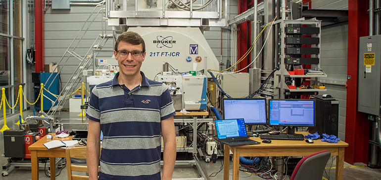
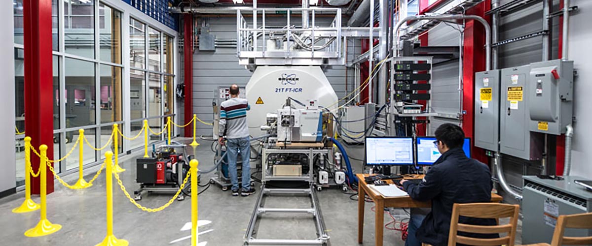
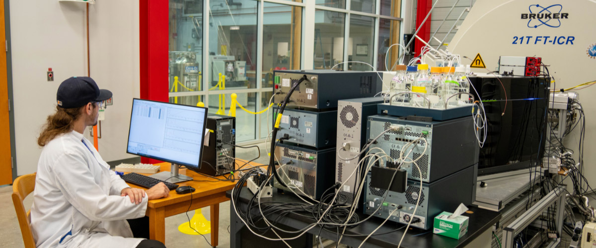
Don Smith at the world-record 21-tesla ICR magnet.
"The 21-T makes this an easy experiment to do," Smith added. "With other mass spectrometers or at lower fields, it can be extremely difficult, or even impossible."
“This is the reason we come to the MagLab,” Heeren said, “to push imaging boundaries and see molecular detail that would otherwise remain hidden.”
The 21-T has proven to be a remarkably versatile instrument, said Chris Hendrickson, director of the lab's ICR facility and a co-author on the paper. "The experiments it has enabled have run the gamut from medical biology to emerging environmental contaminants."
Smith said this technique could become a powerful tool for health research. Currently the 21-T is routinely used to examine the molecular makeup of, among other types of samples, complex proteins. Future MALDI experiments could reveal not just which molecules are in there, but precisely where in a tissue sample each is located.
Cancer researchers could use the technique to examine, on the molecular level, exactly where and how a drug travels through diseased tissue; another scientist could study how an organism responds to exposure to a pollutant. Researchers may even be able to compare examples of the same kind of cell to one another to detect subtle molecular differences. Scientists have been applying to do experiments on the magnet, which is available to them at no cost, on these and other topics.
As for Smith, he and his team (he is hiring a postdoctoral associate to focus on high-resolution MALDI MSI and has acquired new MALDI instrumentation) have their work cut out for them, with plenty of data awaiting analysis.
"We basically tried a little bit of everything," said Smith of last year's experiments.
That's two data sets down, 26 to go.
In addition to Smith, Heeren and Hendrickson, contributors to the article included Andrew Bowman (first author) and Shane Ellis of Maastricht University and National MagLab scientist Greg Blakney. The MALDI instrumentation used for the experiments was made available with support from the Dutch province of Limburg.
Story by Kristen Coyne