
Who is the scientist?
Michael McMahon, an associate professor of radiology at Johns Hopkins School of Medicine, has spent over 15 years studying chemical exchange saturation transfer (CEST), a mechanism for improving the contrast and detail of some magnetic resonance imaging (MRI) scans. CEST has the potential to improve detection of disease or injury over conventional magnetic resonance imaging (MRI).
What is going in the magnet?
Short Answer: A mouse.
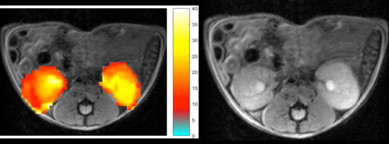
At right is an MRI image of healthy mouse kidneys (circled in red). At left, the same image is overlaid with CEST contrast obtained after the mouse was injected with creatine or phosphocreatine. The yellower areas indicate a higher concentration of the injected compound. Scientists can contrast these CEST images with others taken at different times or with diseased animals to visualize kidney function and detect kidney disease.
Longer Answer: McMahon, an affiliated scientist at the F.M. Kirby Center for Functional Brain Imaging in the Kennedy Krieger Institute, is interested in developing new ways to monitor kidney function. Although techniques such as nuclear imaging (which requires injecting radioactive tracers) can be used for this purpose, they have drawbacks. McMahon had an idea of using MRI (including the strongest MRI machine in the world, located in the MagLab's NMR/MRI Facility) to monitor kidney function, and is performing a series of experiments at the MagLab to test this idea.

Michael McMahon
McMahon thinks, using CEST, that he can use MRI machines to watch how well (or poorly) a kidney functions. Specifically, he thinks he will be able to see signs of two compounds — creatine and phosphocreatine — combining into a third compound called creatinine. This process is a sign of a healthy kidney.
An MRI machine uses a superconducting magnet and specific radio frequencies to map the hydrogen in the water molecules of the body. The hydrogen emits signals that get translated into images scientists use to distinguish different types of soft tissues in the body. This helps doctors locate tumors or view how parts of the body function.
CEST is an emerging technology that allows scientists to see structures or processes in the body that traditional MRI can’t illuminate. Through a rapid “chemical exchange” process, the hydrogen atoms from the molecules the scientist wants to see interchange with hydrogen atoms in water in the body, making those target molecules stand out in MRI scans.
In this case, the molecules of interest are involved in creatine metabolism. McMahon predicts that the MRI scans of a well-functioning kidney will show the levels of creatine and phosphocreatine decrease over time as they are metabolized into creatinine, while creatinine levels increase.
Why do I care?
Each year, chronic kidney disease (CKD) affects more people than breast cancer or prostate cancer. Kidney disease can progress quickly without obvious symptoms, so technologies that allow early detection of damage are essential. McMahon’s research could potentially extend the lives of the millions of people who suffer from CKD by making the diagnostic process more reliable and efficient.
Story By Alexandria Curtwright