
All engineers who design magnets face the same trade-off: power vs. space. The wider the bore (the space inside the magnet where the experiments take place), the weaker the magnetic field inside the bore. While the bore of the typical hospital Magnetic Resonance Imaging (MRI) scanner (a type of NMR magnet) accommodates a human, it produces a field of somewhere between 0.1 and 3 tesla. That's exponentially stronger than a fridge magnet, but much weaker than the fields produced in NMR magnets used for biomedical research.
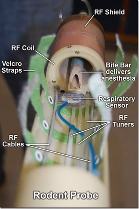
Rodent probe
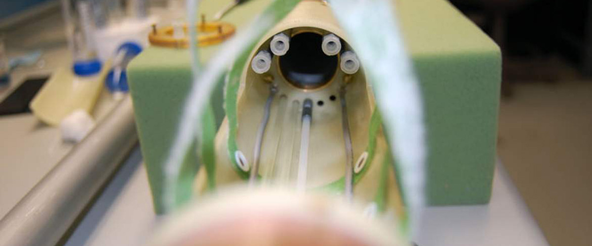
The bore of the MagLab's 900 MHz NMR magnet is only as wide as an orange. But what it lacks in space, it makes up for in power — a magnetic field of 21.1 tesla. Though narrow, the bore is wide enough to accommodate our 33-millimeter (1.3-inch) diameter "rat probe." Scientists can place live rodents in this probe, insert it into the magnet, and create amazing images of these mammals, far more detailed than currently can be obtained of human beings. In essence, the probe turns the NMR magnet into the strongest MRI scanner in the world.
"This is a remarkable step ahead," says Victor Schepkin, a MagLab biophysicist specializing in cancer research. "It's expanding our MR imaging capabilities in sensitivity and resolution. It's increasing the scope of animal models which can be studied at this world-record high magnetic field."
This probe is not the first to allow scientists to examine live animals in a high-field magnet. But it's the first to make possible such experiments in a machine as powerful as the 900 MHz, and with animals as large as adult rats. This magnet is vertical rather than horizontal, as most imaging machines are, and the probe had to be designed around this unusual orientation.
Thanks to this probe, in use at the lab since 2008, Schepkin has been able to image the brains of live rats with amazing clarity, gathering data on brain tumors, traumatic brain injuries and their underlying biology. The ability to study live rats yields a multitude of information impossible to glean otherwise, and means fewer animals are sacrificed.
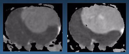
Images of a rat brain produced with the rodent probe used in the 900 MHz NMR magnet.
The probe was developed by MagLab engineer Peter Gor'kov in collaboration with the animal probe team at the lab's Advanced Magnetic Resonance Imaging and Spectroscopy program at the University of Florida in Gainesville. This probe is significant because it allows scientists to move up a notch on the size scale of in vivo experiments, from mouse to rat. The larger an animal's brain, the larger the range of biologically relevant processes inside that brain that can be investigated by MRI. And the stronger the magnet used to collect the magnetic resonance images, the more detailed and useful the images.
Schepkin has been using the probe to study how brain tumors respond to chemotherapy. The images below, obtained using the probe, depict water diffusion in the brain of a rat with a brain tumor.
The image on the left shows the brain prior to chemotherapy; the lighter region is the tumor. The image on the right shows the same part of the brain four days after chemotherapy. The lighter shading in the tumor on the right indicates more water is flowing in those tumor cells, a sign they are breaking down and that the treatment is working.
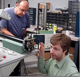
Richard Desilets and Peter Gor'kov work on a probe.
"This is the very first sign that the treatment is working and that the tumor will shrink in the nearest future," says Schepkin.
Packing into the narrow probe all the equipment needed to image a live rat took a lot of crafty planning. First, the designers had to fit in the components needed to create a magnetic resonance image — two radio frequency (RF) cables to transmit and receive signals and four RF tuners to adjust the imaging coils remotely. In addition, they had to include life support equipment to ensure the well-being of the animal: a thermometer, respiratory monitor, climate control, EKG monitor and a way to deliver anesthesia.
Using the probe with live birds, mice and rats, scientists are studying basic biological processes that will help us understand neurodegenerative diseases and other conditions in humans.
Gor'kov, who has designed and built more than 25 specialized probes in his MagLab career, is low-key about the feat. "It's my specialty," he shrugs.
For more information, please contact Public Affairs.