"Darn this aching back!”
That about sums up your thoughts as, flat on your back, clad in a thin hospital gown, you are guided into a dark, tunnel-like tube, a kind of medical solitary confinement where you’ll be spending the next 30 to 60 minutes. Your ears are stuffed with plugs, sound-cancelling headphones cradle your cranium, your extremities are stripped of watch, jewelry and other sentimental comforts, and you may feel a touch of claustrophobia. And it’s all because your orthopedist, worried about that chronic ache in your back, ordered an MRI scan.
Look on the bright side: MRI scans sure beat surgery, which was your only option 20-some years ago, before MRI became a routine, highly-valued diagnostic tool for doctors. In fact, when you take time to think about it, these machines are pretty amazing. And because you’ll be a captive audience while the technologist in the next room takes a bunch of fancy photos of your spine, we suggest you relax and let us tell you the story of the remarkable contraption in which you are, for the time being at least, stuck. We suggest you think of the cylinder as a kind of spacesuit, the machine as a spaceship, and you as an astronaut about to embark on a journey to the center of ... your body!

You probably already have a vague idea of what’s going on. MRI – doesn’t that stand for Magnetic Resonance Imaging? Bull’s-eye. But what does that mean, exactly? Some type of X-ray, isn’t it?
Close, but not quite. MRI scanners, like X-rays and CT scanners, are basically machines doctors use to take pictures of your insides so that they can figure out what’s ailing you. But MRI doesn’t involve ionizing radiation, as do X-rays and CT scans. Rather, MRI takes advantage of something you have plenty of in your body: water. It is far more flexible than X-rays and CT scans, and can generate three dimensional images in any orientation and at any depth in the body.
Physics Factoid
In research and industry, MRI is known as NMR – nuclear magnetic resonance. It's more or less the same process, but the medical establishment prefers the term MRI because some patients are scared off by the word nuclear.
The upshot is that MRI, for most applications, is far superior to other imaging tools in providing non-invasive images (and even chemical information) at high resolution. That’s why hospitals pay millions of dollars for the multi-ton behemoths, and spend hundreds of thousands more a year operating them. Since 1977, when the first MRI exam was conducted on a person, the procedure has become quite common. In 2003, some 60 million scans were performed using about 10,000 MRI scanners worldwide.
While X-rays remain useful for looking at bones, MRI scans are the diagnostic tool of choice for soft tissue – organs, ligaments, the circulatory system and (as you know) the spinal column and cord. They help physicians identify multiple sclerosis, tumors, tendonitis, strokes and many other conditions. What’s more, MRI technology is still in its infancy. Manufacturers are constantly improving scanner designs, and scientists are discovering new applications, from monitoring wine quality to detecting lies; one MRI study revealed that people used twice as many regions of the brain to tell lies as they did to tell the truth.
We know a lot about MRIs at the MagLab because we have the world's strongest MRI scanner: Our 900 MHz nuclear magnetic resonance magnet produces a magnetic field of 21.1 tesla. As you'll soon read, that's far stronger than a hospital MRI scanner. We can't fit humans in it, just laboratory animals such as mice and rats. But the MRI research done on those animals is helping scientists understand a wide variety of human disorders, from Alzheimer's to cancer to muscle degeneration.
We have a lot of ground to cover in describing how this works. In the pages ahead, we’ll learn about the secret lives of hydrogen atoms, how radio waves can make you flip, and facts about superconductivity that will send shivers down your sore spine. It’s all fascinating, but try not to get too excited – you’ll blur your MRI scans!
Magnets with Muscle
Let’s start with a little tour of the metallic cylinder surrounding you (it’s called a bore, technically).
You’re in the center of a tremendous magnet, weighing tens of thousands of pounds and differing from the little magnets on your fridge in two fundamental ways. First, those fridge decorations are permanent magnets made of alloys. The MRI magnet surrounding you, on the other hand, is a superconducting magnet; it conducts electricity, thereby creating a magnetic field. Secondly, your fridge magnet has a fraction of the power of the one you’re in. Scientists measure magnetic strength in units called tesla and gauss – 1 tesla equals 10,000 gauss. The Earth’s magnetic pull is about .5 gauss. Your fridge magnet is about 10 gauss. The electromagnet you’re inside could be up to 3 tesla – 60,000 times the force of the Earth’s magnetic field.
Physics Factoid
The first MRI on a human was made in July 1977 by Dr. Raymond Damadian of New York.
Let’s take a moment to appreciate the “superconducting” part of that magnet, without which your MRI scanner would not be here. You could (and people do) make a permanent magnet with the strength to run an MRI. For the most part, however, these magnets are prohibitively huge and heavy. That leaves you the option of creating a magnet by running electrical current through wire coils – an electromagnet. The problem is the electrons making up that current are forever bumping into the fidgety atomic particles of the material through which they are traveling, slowing them down considerably. (Brush up with a quick review of electricity at this atomic level, if you need to). Given the resistance the current encounters, providing the vast amount of power required to overcome it and generate a magnetic field sufficient to operate an MRI would be prohibitively expensive.
This is where our hero, superconductivity, saves the day! Take special coils and surround them with something really, really cold – liquid helium, at 452.4 degrees below zero on the Fahrenheit scale, does quite nicely. The result? Those over-caffeinated atoms in the conducting wire are frozen into submission. Slowed to a virtual halt, they allow the current to sail right through the miles of wires snaking through an MRI scanner. This technology allows for the construction of hugely powerful magnets like the one surrounding you right now. Most clinical MRI scanners use superconducting magnets. If you’re interested in learning more about this, you can read a more in-depth overview of superconductivity.
By the way, don’t let that little business about liquid helium worry you. It’s insulated in a vacuum, so you won’t need your parka. It wouldn’t be allowed, anyway; zippers, snaps, jewelry and other metals can become life-threatening projectiles in the vicinity of a magnet as powerful as this one. That’s why technicians are very careful to keep metals outside the exam room, and why people with pacemakers and aneurism clips can’t have MRI scans.
By now the technologist in the control room, who is talking you through the exam via an intercom, has started to place you into the main magnetic field in your scanner. The field is running horizontally through the bore from your head to your toes (or vice versa, depending on your position). Because your spine is what she’s interested in, you’re being positioned so that part of your back is in the middle (or isocenter) of the field.
You’d think that being in the middle of such a powerful force would make you feel different – tingly or something. It doesn’t. However, on an atomic level, it’s quite a different story, which takes us from the “M” of MRI to the “R” – Resonance. We’ll understand this better after first taking a close look at the molecules in your body.
Spin Control
You’re made up mostly of water, which means a large number of the atoms inside your body are hydrogen atoms. This turns out to be quite fortuitous, because hydrogen atoms happen to be built in such a way that they react dependably to the forces they will be subjected to inside this scanner. The first of these, as you by now know, is a main magnetic field. The second will be pulses of radio waves. But before we talk about those radio waves, let’s get better acquainted with the H of H2O.
In the nucleus of every hydrogen atom is a positively-charged proton that spins (or precesses, scientifically speaking) around an axis, much in the same way as a child’s top. This spinning generates its own tiny magnetic field, giving the proton its own north and south poles. Now, the nuclei of other atoms spin, too, but for a number of reasons (including, as we’ve mentioned, their sheer quantity), MRI is generally interested only in hydrogen atoms.
Under normal circumstances, these hydrogen protons spin about willy-nilly, on randomly oriented axes, as in the depiction below (more or less), showing hydrogen atoms before the MRI’s magnetic field is turned on.
However, when these atoms are placed in a more powerful magnetic field, it’s as if a drill sergeant blew a whistle: the protons line up at attention. Specifically, the axes realign with the more powerful magnetic field: Half of them face in the direction of the field, the other half in the opposite direction. In the interactive animation above, click in the little box to turn the magnetic field on, and watch what happens. See the Pulse button? Don't touch that dial! We'll get to that in a minute.
Physics Factoid
In recent years, "open MRIs" have become increasingly available, offering more room for claustrophobic and large patients. Some radiologists caution the images may not be as good, as the machines often use weaker magnets.
Did we say half of the protons line up one way, and half the other? Well, not exactly. More precisely, a few more atoms (represented by the little blue guys) line up with the magnetic field, in the low-energy configuration, than in the opposite configuration, which requires a bit more energy. (When we say “few,” we mean, in an MRI powered by a 1.5 tesla magnet, a measly 9 out of 2 million protons!) Those few “leftover” protons (the ones not cancelled out by a proton lined up in the opposite direction) are the ones your MRI scanner will be using. Think of these protons as the wallflowers left on the sidelines at a school dance after everyone else has paired up.
The Wonder of Waves
Now that the magnet has gotten the hydrogen protons lined up at attention, the scanner is ready to subject them to the next step, the one that will result in an actual signal.
You’ve probably been wondering about that coil the technologist placed under your back. No, that’s not one of the main magnetic coils (those are all inside the cylinder, beyond your view). Essentially, it’s a radio transceiver, also called an RF coil, which can communicate with your hydrogen atoms via radio frequency (RF) waves. These waves are close in frequency to those of your favorite FM station. In fact, the room in which the MRI scanner is located is probably shielded so that the local easy listening station doesn't interfere with your images.
Physics Factoid
The vast benefits of MRI have not gone unnoticed by the folks in Oslo who dole out Nobel Prizes. In 2003, Paul C. Lauterbur and Peter Mansfield were jointly awarded the Nobel Prize in Physiology or Medicine for their discoveries related to the technology.
Your technologist is using that coil to send RF pulses at your spine. They are precisely timed (taking into account the type of tissue targeted and the fact that just the hydrogen atoms are of interest) to achieve the effect we’re about to describe. This, by the way, explains the “Resonance” part of MRI (told you we’d get to that!).
Remember those “unmatched” hydrogen protons – the ones (still depicted in blue, in the interactive animation above) left hanging without a partner after the magnetic field caused them all to jump into alignment? Well, those protons absorb the energy of the RF pulses, which causes them to flip on their axes – still in line with the magnetic field, but now in the opposite direction, in the high-energy configuration. Scroll back to the interactive animation and click the Pulse button to see how that works (make sure the magnetic field is still on). See how the unmatched protons flip as the RF pulses (denoted in red) are turned on?
Physics Factoid
MRI patients are sometimes injected with gadolinium, a contrast agent that can make abnormalities such as tumors clearer due to the element's special magnetic properties.
It’s impressive enough that scientists figured out exactly how to make those hydrogen protons do that (the frequency needed is called the Larmor frequency). Now, the real magic happens: When the RF pulse stops, the protons release that absorbed energy, return to their previous alignments and, in so doing, emit a signal back to the coil (also in red, in the opposite direction). You may have noticed that has already happened in the interactive animation. If you missed it, hit the “Pulse” button again for another look.
The signal gets turned into an electric current, which the scanner digitizes. The lower the water content in an area, the fewer hydrogen protons there will be emitting signals back to the RF coils. Different types of MRIs display this data differently, but in any case you get a variety of shades of grey that reflect the different densities. In some scans, the weaker the signal, the darker that part of the image will be. So bone will be fairly dark, while fat will be light.
Slicing and Dicing
What’s that? You need me to repeat that last part? I know – it can be hard to hear anything over the periodic hammering sound the scanner generates. Which reminds me – I’ve forgotten to mention anything about what’s making all that noise (and why you’re thankful for those earplugs).

Physics Factoid
MRI patients are sometimes injected with gadolinium, a contrast agent that can make abnormalities such as tumors clearer due to the element's special magnetic properties.
Responsible for that racket are the gradient magnets. There are three of them in the scanner (called x, y and z), each oriented along a different plane of your body, all of them far less powerful than the main magnet. But what they lack in strength they make up for in precision. They modify the magnetic field at very particular points and work in conjunction with the RF pulses to produce the scanner’s picture by encoding the spatial distribution of the water protons in your body. When rapidly turned on and off (which causes that banging noise), the gradient magnets allow the scanner to image the body in slices – sort of like a loaf of bread. Using medical terminology, the transverse (or axial, or x-y) planes slice you from top to bottom; the coronal (x-z) plane slice you lengthwise from front to back; and the sagittal (y-z) planes slice you lengthwise from side to side. However, the x, y and z gradients can be used in combination to generate image slices that are in any direction, which is one of the great strengths of MRI as a diagnostic tool.

Starting to feel like chopped liver?
Let’s put a rush order on your MRI, so that you can see what we mean.
Shades of Gray
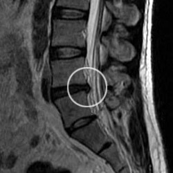
Herniated disc
Here’s a picture (sagittal view) of your spine! Now it’s clear what the trouble is. See the dark disc that, unlike the others, protrudes into the spinal canal? That’s a herniated disc compressing the nerves of the spinal cord. Ouch!
Believe it or not, MRI scans can display more than 250 distinct shades of grey, each reflecting slight variations in tissue density or water content. It is in those subtle shades that radiologists unlock the secrets of the tissues. For example, abnormal tissue, such as a brain tumor, will look different than the normal tissue surrounding it. The technologist and radiologist have the ability to alter imaging parameters (like the timings of the RF pulse and gradients) to emphasize areas of injury or disease or to acquire higher image resolutions.
Now that your scan is over, you’re free to move around – do jumping jacks, dance a jig, celebrate your liberation. Unstop your ears, trade in the hospital gown for your street duds, reclaim your jewelry and watch. The MRI has not changed anything about your body or its chemistry. In fact, unlike X-rays or CT scans, you can have as many MRI scans as often as is necessary to diagnose your ailment and track your recovery after treatment.
Physics Factoid
MRIs are most commonly used for cancer patients (about 35 percent of all scans) and patients with spinal problems (about 30 percent).
We hope that the bad news about your back has been tempered by a newfound appreciation for the beauties of science and the wonders of technology. If we’ve whetted your appetite for more information on the topic, you might visit some of the Web sites listed below.
For a more complete explanation of how MRI works, we invite you to view an interactive tutorial about Magnetic Resonance Imaging (MRI) and watch a video about How MRI Machines Work.
Thanks to our Science Adviser for this article: Sam Grant, an assistant professor of engineering with the Florida A&M University/Florida State University College of Engineering and an expert in MRI at the MagLab.
By Kristen Coyne